
It has been proven by scientific studies that a sedentary lifestyle is not compatible with human gene structure. It is known that the less active lifestyle that has emerged due to technological developments is an important factor in the formation of many chronic diseases.
Weight, diet and exercise directly affect human health and cancer risk. There are studies showing that the risk of uterine, lung and prostate cancers also decreases with exercise. Recent studies and the American Centers for Disease Control and Prevention (CDC) recommend at least 30 minutes of moderate-to-vigorous exercise 3 or 5 days a week to reduce the risk of cancer. Accordingly, regular exercise prevents the development of many types of cancer, especially breast cancer.
Most recent studies show that women who exercise have a lower risk of developing breast cancer than those who do not. Most of the data show that exercise reduces the risk of breast cancer in both premenopausal and postmenopausal women. Moderate to vigorous exercise during adolescence provides a particularly protective effect. Although lifelong regular and vigorous exercise is thought to be the most beneficial, women who increase their exercise even after menopause have a reduced risk compared to women who do not exercise.
According to some studies, the effect of exercise varies according to BMI (Body Mass Index). The benefit of exercise is greatest in those with a BMI below 25, that is, those whose weight is in the normal range. The data show that as the duration and frequency of exercise increases, the risk of breast cancer decreases. Most studies show that 30-60 minutes/day of moderate to vigorous physical activity reduces the risk of breast cancer.
Exercise prevents tumor development by lowering hormone levels, especially in premenopausal women. Exercise lowers insulin and insulin-like growth factor (IGF-1) levels in the blood, increases immune response, and prevents excess body fat and high body mass by maintaining ideal weight.
Studies show that exercise can be beneficial in increasing the quality of life, reducing fatigue and establishing energy balance after the diagnosis of breast cancer. Often, after a breast cancer diagnosis, both treatment and reduced movement lead to weight gain. However, a woman who does moderate exercise after a breast cancer diagnosis may have a longer survival than a woman who is sedentary. This benefit is even more pronounced in the hormone-sensitive patient.
Regular exercise for 4-5 hours a week alleviates the side effects of radiation and drug treatments, increases psychological well-being and reduces the risk of recurrence of the disease, even in patients with breast cancer.
The estrogen hormone secreted by the fat cells in the body is the most important risk factor for the formation of breast cancer. The fact that the fat ratio is lower in people who do sports regularly will reduce the estrogen level, thus playing a role in preventing the formation of breast cancer.
In addition, insulin-like growth factors (IGF) in the blood increase the risk of breast cancer by stimulating the division of breast cells. Regular exercise reduces the risk of developing breast cancer by preventing the increase in insulin and IGF in the blood.
Due to the intensity of work and family life, it may seem impossible to allocate time to exercise, as well as to do it regularly and continuously. However, daily exercise times can be increased with some arrangements. Walking is a good choice. You can walk for half an hour before going to work or during lunch breaks. If you are driving, parking far away from your work and home will increase your daily walking time. Taking a walk with a friend after work will both increase your exercise rate and help you relieve the stress of the day.
You can use gyms for different types of exercise, you can work out at home with exercise videos or dance to music. If you choose the exercise method that does not bore you and best suits your daily life routine, it will be easier to maintain it regularly.
As a result, it is clearly seen that exercise has an important role in the prevention and treatment of cancer. Being physically active throughout life reduces the risk of many cancers, especially colon-rectum and breast cancer. For this reason, regular sports should be practiced especially by women at high risk for breast cancer.
Meme hastalıkları ve tedavisi konusunda eğitimi olan doktorlar onkolojik ve onkoplastik meme cerrahisi uzmanlarıdır. Memenin klinik muayenesi için, genel cerrahi uzmanına, daha ideal olarak, genel cerrahlar içinden meme hastalıkları ve cerrahisi konusunda uzmanlaşmış bir onkolojik ve onkoplastik meme cerrahına gitmek en uygun olanıdır.
Meme hastalıkları, meme kanseri tedavisi ve meme cerrahisi bilgi ve deneyim gerektiren konulardır. Özel olarak meme hastalıkları ve tedavisi ile ilgilenen genel cerrahi uzmanları yani meme cerrahları memenin klinik muayenesi için seçilecek doktorlar olmalıdır.
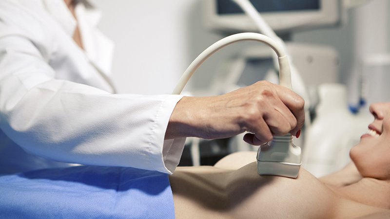
Memede kitle fark edildiğin ilk olarak genel cerrahi uzmanından randevu alınması gerekiyor. İlk görüşmede bazı bilgiler isteniyor hastadan. Risk faktörleri değerlendiriliyor, geçmiş öyküsü alınıyor, kullandığı ilaçlar soruluyor. Ardından muayene ediliyor hasta. Muayenede meme ve koltuk altında kitle hangi bölgede, deri ile ilişkisi nasıl, deride çekintiye sebep oluyor mu? Koltuk altında lenf bezi var mı? Bunlar kontrol ediliyor. Daha sonra hastanın yaşına göre mamografi ya da ultrason tetkiki yapılıyor.
Genelde 40 yaş sonrası için mamografi öneriliyor. Mamografi düşük dozda X ışını içeren bir tetkik. Genellikle dijital mamografi tercih ediliyor. Ultrason ise mamografiye yardımcı olarak genellikle genç hastalarda yol gösterici oluyor. Hasta 35 yaş altı ise öncellikle ultrasona başvuruluyor. Ultrason X ışını değil de ses dalgası yöntemine göre çalışıyor. Kitle kist mi, içinde sıvı var mı yoksa solid mi katı mı buna bakılıyor. Sonraki aşamada ise katı içerikli kitlelere biyopsi yapılıyor. Biyopsi için birkaç yöntem var, bunlardan bir tanesi ince iğne – kalın iğne biyopsisi ve cerrahi biyopsi. Cerrahi biyopsi günümüzde çok tercih edilmiyor, daha çok kalın iğne biyopsisi yapılıyor. Kalın iğne biyopsisi nasıl yapılıyor? Bölgenin uyuşturulması sonrası otomatik bir tabancayla birkaç örnek alınıyor ve patoloji incelemesine gönderiliyor. Bu inceleme bize kitle hakkında bilgi veriyor. Eğer tümör hücresi içerisiyorsa biyopsi sonucuna ek incelemeler de yapılıyor. İmmün testler yapılıyor. Bu testlerde östrojen ve progesteron reseptörleri, her 2 düzeyi gibi bilgilere ihtiyaç oluyor. Daha sonraki aşamada hastanın evresine göre ek taramalar yapılabiliyor. Bu aşamada sıklıkla PET-CT öneriliyor. Hem sonuca kolay ulaşılıyor hem de tek tetkikte birçok organ gözlenebiliyor. PET-CT çekildikten sonra tedavi planı yapılıyor. Erken dönemdeki hastalarda daha çok cerrahi ile başlanıyor, ileri evre hastalarda ise öncelikle sistemik tedavi denilen kemoterapi ya da hormon ilaçlarıyla başlıyoruz.
Meme cerrahının veya genel cerrahi uzmanının, hastanın meme ile ilgili risklerini sorgulayarak, meme muayenesi yapmasına klinik meme muayenesi denir.
Memede kanser, 8 kadında bir görülen, görülme sıklığı kadınlarda ilk sırada olan bir kanser türüdür. Meme de kitle ise toplumda 4 kadında 1 kadında görülür.
Meme cerrahı veya genel cerrahi uzmanı klinik meme muayenesi ile hastanın meme kanseri için risk yaratan durumlarını, hormonal durumunu, aile hikayesini, hastalıklarını, doğurganlığını, emzirme veya vücut kitle indeksi (BMI) gibi kişisel durumlarını, meme sağlığını etkileyebilecek ilaçlarını ve alışkanlıklarını, meme ile ilgili daha önce geçirdiği hastalıkları, ameliyat veya biyopsileri sorgulayarak memeyi muayene eder.
Tüm bu sorgulamalardan elde ettiği verileri, hastanın elle yapılan muayenesi ile birleştirir. Hastanın elde olan verilerine uygun görüntüleme yöntemi veya yöntemlerinin önerir. Bu görüntüleme sonucunda elde edilen verileri, klinikte elde ettiği muayene ve sorgulama verileriyle birleştirir.
Gerekiyorsa daha ileri ek görüntülemeler ve tetkikler ister. Sonuçta meme cerrahı veya genel cerrahi uzmanı, her hasta için bir meme kanseri risk değerlendirmesi ile hastaya özel kişiselleştirilmiş izlem önerir.

Meme muayenesi, öncelikli olarak meme hastalıkları ve meme kanseri konusunda risk yaratacak durumlara neden olacak konuları sorgulayarak başlamalıdır.
Klinik meme muayenesine, hastanın memesindeki yakınması ile ilgili durumları etkileyecek tüm olasılıklar sorgulanarak başlanır.
Meme hastalıkları konusunda yakınması olan hastada, geçirdiği ameliyatlar, aile hikayesi, hormonal düzeni, kullandığı ilaçlar, mesleği, alışkanlıkları, evlilik ve doğum durumu, emzirme, genetik yatkınlığı, beraberinde olan hastalıkları ve kullandığı veya geçirdiği tedaviler ayrıntılı olarak sorgulanır.
Hastanın bu durumla ilgili var olan geçmiş zamana ait tüm tetkikleri değerlendirilir. Sonra meme cerrahı veya genel cerrahi uzmanı memede ağrı, memede kitle, memede enfeksiyon, memede kist ve benzeri yakınması olan kişiyi elle muayene eder. Bu elle meme muayenesi, memenin kapladığı tüm alanı, memedeki yakınmalara neden olacak yansıması olacak alanları, olası meme kanserinin yayılabileceği / sıçrayabileceği tüm alanları ve lenf bezlerinin klinik elle muayenesini içerir.
Elle meme muayenesinden elde edilen tüm bulgular ve özellikleri kaydedilir. Hastanın sorgulamasından elde edilen verilerle birleştirilerek değerlendirilir.
Meme konusunda yakınması olan hasta için, yakınmasına ve yaşına en uygun görüntüleme ve gerekli kan tetkikleri istenir. Daha sonra meme cerrahı bu meme görüntüleme ve kan tetkiki sonuçlarını hastanın klinik meme muayenesi ve yakınmalarına göre yorumlar.
Elde edilen sonuca göre ya daha ileri tetkiklere geçilir. Ya da saptanan duruma uygun tedavi veya meme hastalığının izlemi önerilir. Belirlenen izlem aralığına göre hastanın, meme klinik muayenesinden elde edilen bulguların zaman içinde değişiklik gösterip göstermediği, ilerleyip ilerlemediği, bir tedavi önerildi ise tedaviye yanıtı değerlendirilir. Bu meme izlemlerinde hastaya uygun radyolojik meme görüntüleme yöntemleri de tekrar edilebilir ve önceki değerlendirmelerle karşılaştırılır.

It has been reported that a substance that is a by-product of cholesterol can trigger the formation and spread of breast cancer. These findings sparked the hope of preventing cancer through taking cholesterol-lowering drugs called statins. The study published in the journal Science (http://www.sciencemag.org/content/342/6162/1094) also explains why obesity is an important factor in cancer. Similar studies have been done before on the relationship between statins and breast cancer. Similar findings were noted in a retrospective study conducted by the University of Texas MD Anderson Cancer Center and presented at the San Antonio Breast Cancer Symposium. 6 months ago, the Medical Academy published a news article about the results of the study in this presentation, under the title “Statins improve survival in inflammatory breast cancer”.
In the report, it was discussed in the light of the information in the presentation that statins, which are widely used to lower cholesterol, improve progression-free survival in patients with inflammatory breast cancer (IBC). We also recommend our readers to take a look at this article. The BBC, which published the study published in Science magazine today (November 29), includes the opinions of the experts who conducted the new study.
“It has been reported that a substance that is a byproduct of cholesterol can trigger the formation and spread of breast cancer. These findings sparked the hope of preventing cancer through taking cholesterol-lowering drugs called statins. But cancer charities warned it was premature to advise women to take statins. The relationship between obesity and breast, colon and uterine cancer has been known for some time.
Fat in overweight people can cause cancer by causing the secretion of hormones such as estrogen. The team conducting the research at Duke University Medical Center in the USA revealed that cholesterol has the same effect. The human body converts cholesterol into a by-product called 27HC, and this substance, which is an imitation of estrogen, shows the effect of this hormone in some tissues. Experiments on mice showed that a high-fat diet increased blood 27HC, and tumors grew 30% larger and were more likely to spread than mice on a normal diet. Breast cancer tissue was also found to grow faster when fed 27HC in the laboratory.
One of the researchers Prof. Dr. Donald McDonnell said that many previous studies have shown the link between obesity and breast cancer and that excess cholesterol increases the risk of breast cancer, but that no specific mechanism has been identified. prof. Dr. “The molecule we’ve now found, called 27HC, which is not cholesterol itself but a byproduct, mimics the estrogen hormone and can trigger breast cancer on its own,” McDonnell said. said.
The researchers say these findings strengthen hopes of lowering the risk of breast cancer through lowering cholesterol. Statin is a substance used by millions of people against heart disease. But studies have also shown that this substance also reduces the risk of breast cancer. Another way to lower the cholesterol level in the blood is through a healthy diet. Dr Emma Smith, from the Cancer Research Foundation UK, said: “This is the first time that this research shows a direct link between cholesterol and breast cancer in mice, but it is too early to talk about how this information will work in the future in the fight against breast cancer. “Until we know more about the impact of statins on cancer risk, the best way to reduce this risk is to stay at a good weight, reduce alcohol consumption and exercise.”

Doctors make every effort to ensure that women with breast cancer can return to their usual activities as soon as possible. The recovery period varies for each woman, depending on the stage of the disease, the type of treatment and other factors.
After the operation, the woman’s arm and shoulder exercise helps this region regain strength and movement; reduces pain and muscle stiffness in the back and neck. Special exercises usually start a few days after the tubes that are placed under the skin and drained during the surgery and drain the accumulated blood and lymph fluid are removed. Exercises start slowly and for short periods, and can be done even in bed. It is mostly performed under the supervision of an experienced physiotherapist. As time passes, the quality and duration of the exercises can be increased. Regular exercise turns into a woman’s daily routine in the following period.
Women who have had a mastectomy or breast reconstruction should do the specific exercises their doctor will explain.
Doing exercises frequently and placing the arm on the pillow prevents or reduces lymphedema (swelling in the hand, arm and forearm) that may occur after surgery. Information about prevention and treatment of lymphedema is explained in the “Side Effects” section.