It has been proven by scientific studies that a sedentary lifestyle is not compatible with human gene structure. It is known that the less active lifestyle that has emerged due to technological developments is an important factor in the formation of many chronic diseases.
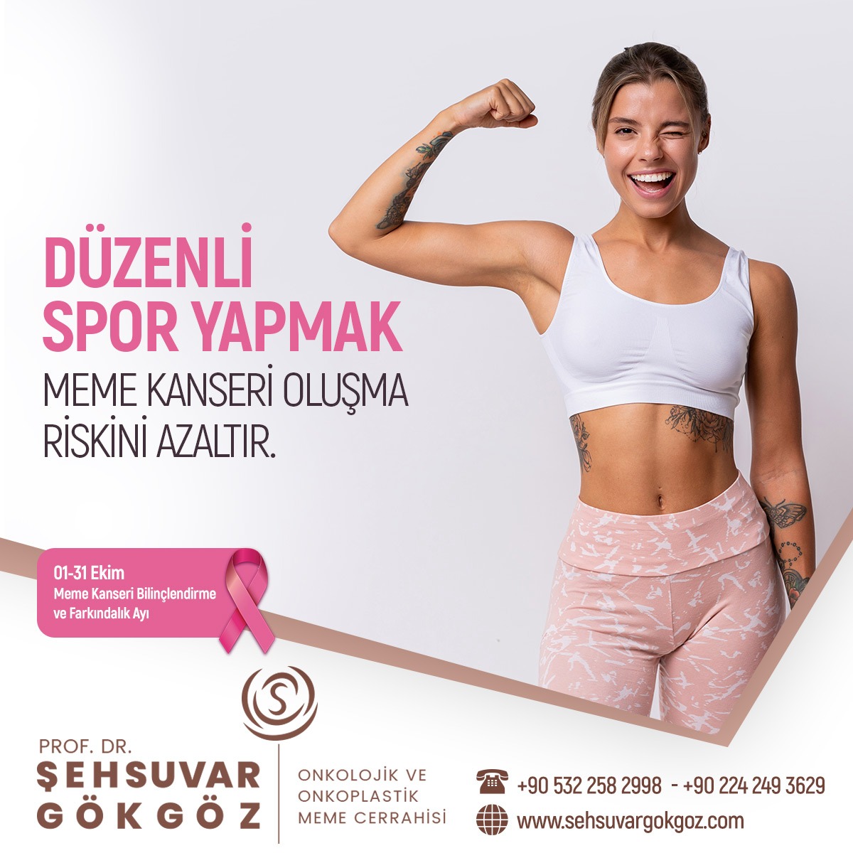
Weight, diet and exercise directly affect human health and cancer risk. There are studies showing that the risk of uterine, lung and prostate cancers also decreases with exercise. Recent studies and the American Centers for Disease Control and Prevention (CDC) recommend at least 30 minutes of moderate-to-vigorous exercise 3 or 5 days a week to reduce the risk of cancer. Accordingly, regular exercise prevents the development of many types of cancer, especially breast cancer.
Most recent studies show that women who exercise have a lower risk of developing breast cancer than those who do not. Most of the data show that exercise reduces the risk of breast cancer in both premenopausal and postmenopausal women. Moderate to vigorous exercise during adolescence provides a particularly protective effect. Although lifelong regular and vigorous exercise is thought to be the most beneficial, women who increase their exercise even after menopause have a reduced risk compared to women who do not exercise.
According to some studies, the effect of exercise varies according to BMI (Body Mass Index). The benefit of exercise is greatest in those with a BMI below 25, that is, those whose weight is in the normal range. The data show that as the duration and frequency of exercise increases, the risk of breast cancer decreases. Most studies show that 30-60 minutes/day of moderate to vigorous physical activity reduces the risk of breast cancer.
Exercise prevents tumor development by lowering hormone levels, especially in premenopausal women. Exercise lowers insulin and insulin-like growth factor (IGF-1) levels in the blood, increases immune response, and prevents excess body fat and high body mass by maintaining ideal weight.
Studies show that exercise can be beneficial in increasing the quality of life, reducing fatigue and establishing energy balance after the diagnosis of breast cancer. Often, after a breast cancer diagnosis, both treatment and reduced movement lead to weight gain. However, a woman who does moderate exercise after a breast cancer diagnosis may have a longer survival than a woman who is sedentary. This benefit is even more pronounced in the hormone-sensitive patient.
Regular exercise for 4-5 hours a week alleviates the side effects of radiation and drug treatments, increases psychological well-being and reduces the risk of recurrence of the disease, even in patients with breast cancer.
The estrogen hormone secreted by the fat cells in the body is the most important risk factor for the formation of breast cancer. The fact that the fat ratio is lower in people who do sports regularly will reduce the estrogen level, thus playing a role in preventing the formation of breast cancer.
In addition, insulin-like growth factors (IGF) in the blood increase the risk of breast cancer by stimulating the division of breast cells. Regular exercise reduces the risk of developing breast cancer by preventing the increase in insulin and IGF in the blood.
Due to the intensity of work and family life, it may seem impossible to allocate time to exercise, as well as to do it regularly and continuously. However, daily exercise times can be increased with some arrangements. Walking is a good choice. You can walk for half an hour before going to work or during lunch breaks. If you are driving, parking far away from your work and home will increase your daily walking time. Taking a walk with a friend after work will both increase your exercise rate and help you relieve the stress of the day.
You can use gyms for different types of exercise, you can work out at home with exercise videos or dance to music. If you choose the exercise method that does not bore you and best suits your daily life routine, it will be easier to maintain it regularly.
As a result, it is clearly seen that exercise has an important role in the prevention and treatment of cancer. Being physically active throughout life reduces the risk of many cancers, especially colon-rectum and breast cancer. For this reason, regular sports should be practiced especially by women at high risk for breast cancer.

With the Contribution of the International Breast Health Working Group (BHWGI)
21-22 Ekim | 2022-ci yıl
Fairmont Bakü, Flame Towers | Bakü, Azerbaycan
T. +994502438671
21- 22 – October 2022 / Baku / Azerbaijan Let’s meet at the 1st International Breast Cancer Congress..
Prof. Dr. M. Şehsuvar GÖKGÖZ
Oncological and Oncoplastic Breast Surgery
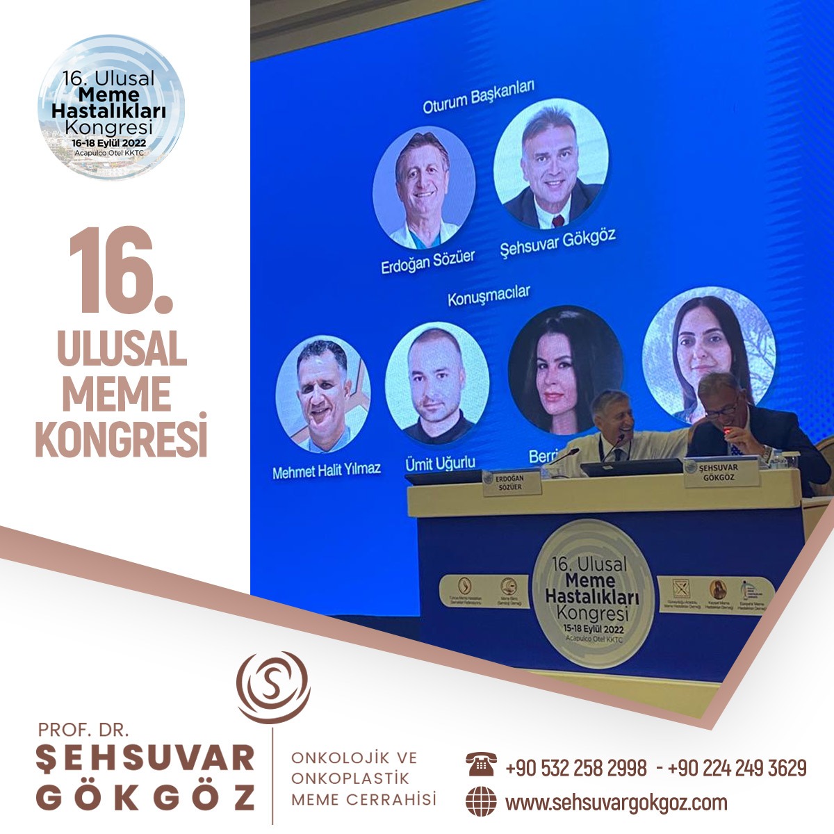
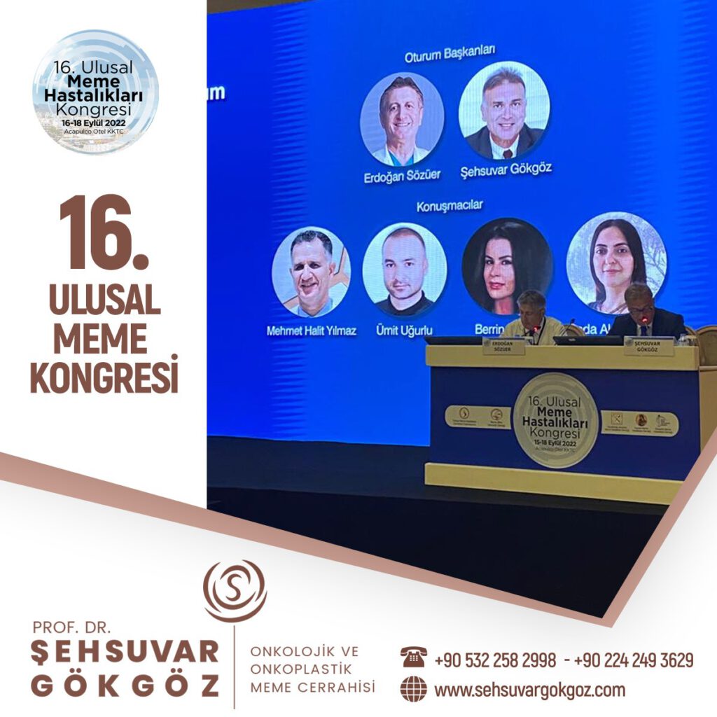
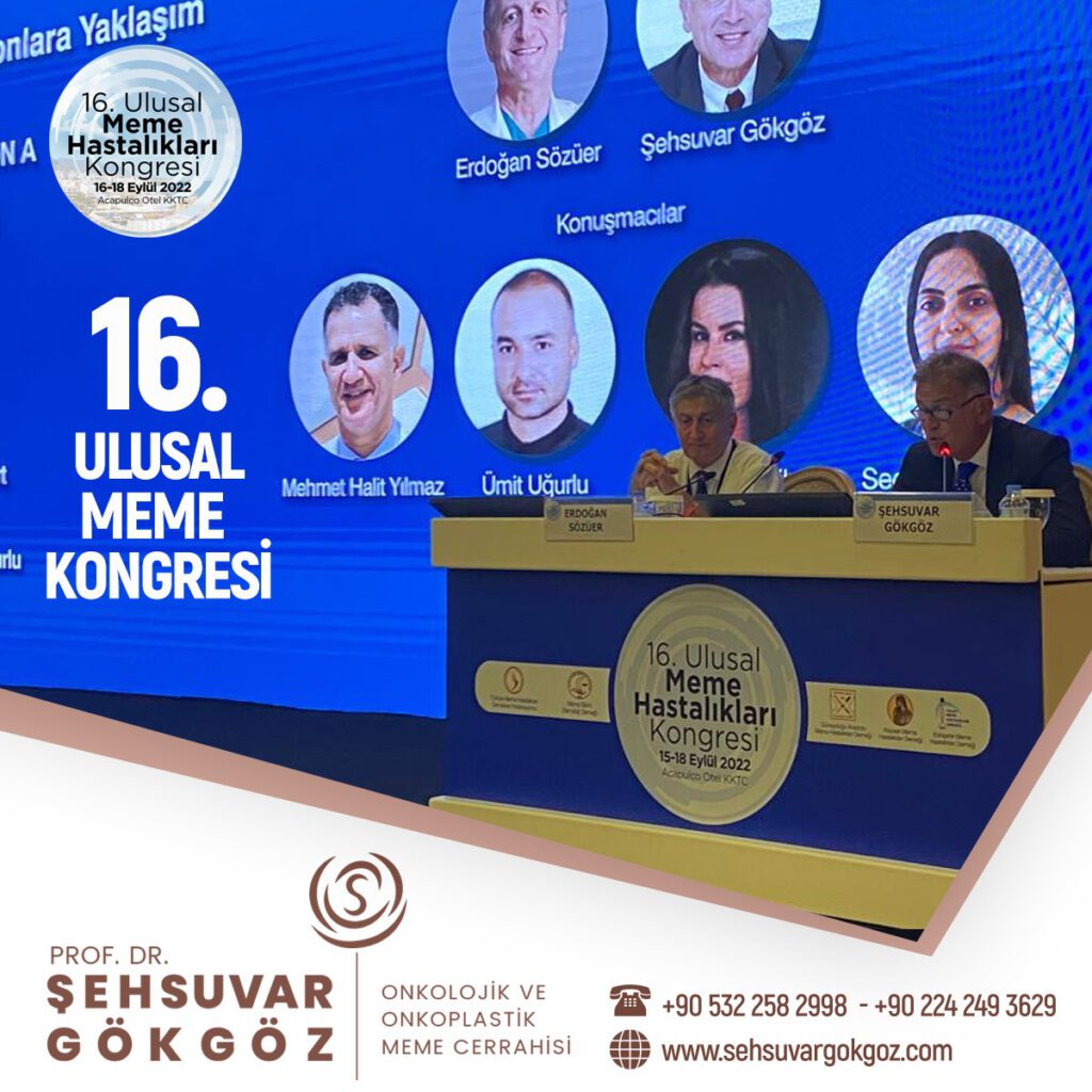
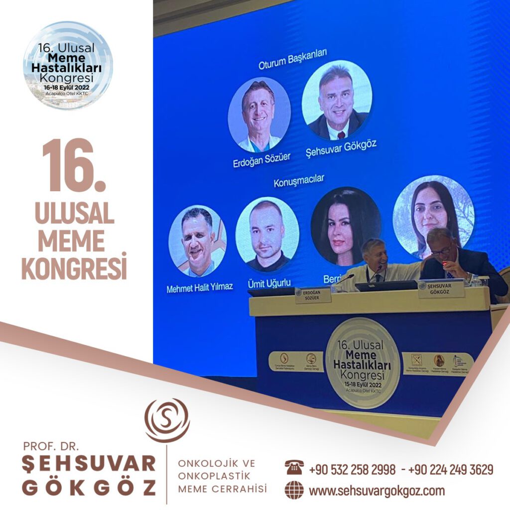
Breast diseases and breast cancer are an increasingly important health problem in our country. In recent years, studies on treatment approaches and options have accelerated. I was at the National Breast Diseases Congress held between 16-18 September in order to discuss these issues in detail and to share current developments with our colleagues.
Meme hastalıkları ve tedavisi konusunda eğitimi olan doktorlar onkolojik ve onkoplastik meme cerrahisi uzmanlarıdır. Memenin klinik muayenesi için, genel cerrahi uzmanına, daha ideal olarak, genel cerrahlar içinden meme hastalıkları ve cerrahisi konusunda uzmanlaşmış bir onkolojik ve onkoplastik meme cerrahına gitmek en uygun olanıdır.
Meme hastalıkları, meme kanseri tedavisi ve meme cerrahisi bilgi ve deneyim gerektiren konulardır. Özel olarak meme hastalıkları ve tedavisi ile ilgilenen genel cerrahi uzmanları yani meme cerrahları memenin klinik muayenesi için seçilecek doktorlar olmalıdır.
Memede kitle fark edildiğin ilk olarak genel cerrahi uzmanından randevu alınması gerekiyor. İlk görüşmede bazı bilgiler isteniyor hastadan. Risk faktörleri değerlendiriliyor, geçmiş öyküsü alınıyor, kullandığı ilaçlar soruluyor. Ardından muayene ediliyor hasta. Muayenede meme ve koltuk altında kitle hangi bölgede, deri ile ilişkisi nasıl, deride çekintiye sebep oluyor mu? Koltuk altında lenf bezi var mı? Bunlar kontrol ediliyor. Daha sonra hastanın yaşına göre mamografi ya da ultrason tetkiki yapılıyor.
Genelde 40 yaş sonrası için mamografi öneriliyor. Mamografi düşük dozda X ışını içeren bir tetkik. Genellikle dijital mamografi tercih ediliyor. Ultrason ise mamografiye yardımcı olarak genellikle genç hastalarda yol gösterici oluyor. Hasta 35 yaş altı ise öncellikle ultrasona başvuruluyor. Ultrason X ışını değil de ses dalgası yöntemine göre çalışıyor. Kitle kist mi, içinde sıvı var mı yoksa solid mi katı mı buna bakılıyor. Sonraki aşamada ise katı içerikli kitlelere biyopsi yapılıyor. Biyopsi için birkaç yöntem var, bunlardan bir tanesi ince iğne – kalın iğne biyopsisi ve cerrahi biyopsi. Cerrahi biyopsi günümüzde çok tercih edilmiyor, daha çok kalın iğne biyopsisi yapılıyor. Kalın iğne biyopsisi nasıl yapılıyor? Bölgenin uyuşturulması sonrası otomatik bir tabancayla birkaç örnek alınıyor ve patoloji incelemesine gönderiliyor. Bu inceleme bize kitle hakkında bilgi veriyor. Eğer tümör hücresi içerisiyorsa biyopsi sonucuna ek incelemeler de yapılıyor. İmmün testler yapılıyor. Bu testlerde östrojen ve progesteron reseptörleri, her 2 düzeyi gibi bilgilere ihtiyaç oluyor. Daha sonraki aşamada hastanın evresine göre ek taramalar yapılabiliyor. Bu aşamada sıklıkla PET-CT öneriliyor. Hem sonuca kolay ulaşılıyor hem de tek tetkikte birçok organ gözlenebiliyor. PET-CT çekildikten sonra tedavi planı yapılıyor. Erken dönemdeki hastalarda daha çok cerrahi ile başlanıyor, ileri evre hastalarda ise öncelikle sistemik tedavi denilen kemoterapi ya da hormon ilaçlarıyla başlıyoruz.
Meme cerrahının veya genel cerrahi uzmanının, hastanın meme ile ilgili risklerini sorgulayarak, meme muayenesi yapmasına klinik meme muayenesi denir.
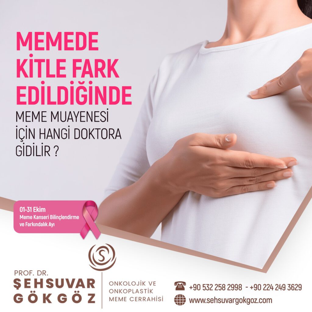
Memede kanser, 8 kadında bir görülen, görülme sıklığı kadınlarda ilk sırada olan bir kanser türüdür. Meme de kitle ise toplumda 4 kadında 1 kadında görülür.
Meme cerrahı veya genel cerrahi uzmanı klinik meme muayenesi ile hastanın meme kanseri için risk yaratan durumlarını, hormonal durumunu, aile hikayesini, hastalıklarını, doğurganlığını, emzirme veya vücut kitle indeksi (BMI) gibi kişisel durumlarını, meme sağlığını etkileyebilecek ilaçlarını ve alışkanlıklarını, meme ile ilgili daha önce geçirdiği hastalıkları, ameliyat veya biyopsileri sorgulayarak memeyi muayene eder.
Tüm bu sorgulamalardan elde ettiği verileri, hastanın elle yapılan muayenesi ile birleştirir. Hastanın elde olan verilerine uygun görüntüleme yöntemi veya yöntemlerinin önerir. Bu görüntüleme sonucunda elde edilen verileri, klinikte elde ettiği muayene ve sorgulama verileriyle birleştirir.
Gerekiyorsa daha ileri ek görüntülemeler ve tetkikler ister. Sonuçta meme cerrahı veya genel cerrahi uzmanı, her hasta için bir meme kanseri risk değerlendirmesi ile hastaya özel kişiselleştirilmiş izlem önerir.

Meme muayenesi, öncelikli olarak meme hastalıkları ve meme kanseri konusunda risk yaratacak durumlara neden olacak konuları sorgulayarak başlamalıdır.
Klinik meme muayenesine, hastanın memesindeki yakınması ile ilgili durumları etkileyecek tüm olasılıklar sorgulanarak başlanır.
Meme hastalıkları konusunda yakınması olan hastada, geçirdiği ameliyatlar, aile hikayesi, hormonal düzeni, kullandığı ilaçlar, mesleği, alışkanlıkları, evlilik ve doğum durumu, emzirme, genetik yatkınlığı, beraberinde olan hastalıkları ve kullandığı veya geçirdiği tedaviler ayrıntılı olarak sorgulanır.
Hastanın bu durumla ilgili var olan geçmiş zamana ait tüm tetkikleri değerlendirilir. Sonra meme cerrahı veya genel cerrahi uzmanı memede ağrı, memede kitle, memede enfeksiyon, memede kist ve benzeri yakınması olan kişiyi elle muayene eder. Bu elle meme muayenesi, memenin kapladığı tüm alanı, memedeki yakınmalara neden olacak yansıması olacak alanları, olası meme kanserinin yayılabileceği / sıçrayabileceği tüm alanları ve lenf bezlerinin klinik elle muayenesini içerir.
Elle meme muayenesinden elde edilen tüm bulgular ve özellikleri kaydedilir. Hastanın sorgulamasından elde edilen verilerle birleştirilerek değerlendirilir.
Meme konusunda yakınması olan hasta için, yakınmasına ve yaşına en uygun görüntüleme ve gerekli kan tetkikleri istenir. Daha sonra meme cerrahı bu meme görüntüleme ve kan tetkiki sonuçlarını hastanın klinik meme muayenesi ve yakınmalarına göre yorumlar.
Elde edilen sonuca göre ya daha ileri tetkiklere geçilir. Ya da saptanan duruma uygun tedavi veya meme hastalığının izlemi önerilir. Belirlenen izlem aralığına göre hastanın, meme klinik muayenesinden elde edilen bulguların zaman içinde değişiklik gösterip göstermediği, ilerleyip ilerlemediği, bir tedavi önerildi ise tedaviye yanıtı değerlendirilir. Bu meme izlemlerinde hastaya uygun radyolojik meme görüntüleme yöntemleri de tekrar edilebilir ve önceki değerlendirmelerle karşılaştırılır.

It has been reported that a substance that is a by-product of cholesterol can trigger the formation and spread of breast cancer. These findings sparked the hope of preventing cancer through taking cholesterol-lowering drugs called statins. The study published in the journal Science (http://www.sciencemag.org/content/342/6162/1094) also explains why obesity is an important factor in cancer. Similar studies have been done before on the relationship between statins and breast cancer. Similar findings were noted in a retrospective study conducted by the University of Texas MD Anderson Cancer Center and presented at the San Antonio Breast Cancer Symposium. 6 months ago, the Medical Academy published a news article about the results of the study in this presentation, under the title “Statins improve survival in inflammatory breast cancer”.
In the report, it was discussed in the light of the information in the presentation that statins, which are widely used to lower cholesterol, improve progression-free survival in patients with inflammatory breast cancer (IBC). We also recommend our readers to take a look at this article. The BBC, which published the study published in Science magazine today (November 29), includes the opinions of the experts who conducted the new study.
“It has been reported that a substance that is a byproduct of cholesterol can trigger the formation and spread of breast cancer. These findings sparked the hope of preventing cancer through taking cholesterol-lowering drugs called statins. But cancer charities warned it was premature to advise women to take statins. The relationship between obesity and breast, colon and uterine cancer has been known for some time.
Fat in overweight people can cause cancer by causing the secretion of hormones such as estrogen. The team conducting the research at Duke University Medical Center in the USA revealed that cholesterol has the same effect. The human body converts cholesterol into a by-product called 27HC, and this substance, which is an imitation of estrogen, shows the effect of this hormone in some tissues. Experiments on mice showed that a high-fat diet increased blood 27HC, and tumors grew 30% larger and were more likely to spread than mice on a normal diet. Breast cancer tissue was also found to grow faster when fed 27HC in the laboratory.
One of the researchers Prof. Dr. Donald McDonnell said that many previous studies have shown the link between obesity and breast cancer and that excess cholesterol increases the risk of breast cancer, but that no specific mechanism has been identified. prof. Dr. “The molecule we’ve now found, called 27HC, which is not cholesterol itself but a byproduct, mimics the estrogen hormone and can trigger breast cancer on its own,” McDonnell said. said.
The researchers say these findings strengthen hopes of lowering the risk of breast cancer through lowering cholesterol. Statin is a substance used by millions of people against heart disease. But studies have also shown that this substance also reduces the risk of breast cancer. Another way to lower the cholesterol level in the blood is through a healthy diet. Dr Emma Smith, from the Cancer Research Foundation UK, said: “This is the first time that this research shows a direct link between cholesterol and breast cancer in mice, but it is too early to talk about how this information will work in the future in the fight against breast cancer. “Until we know more about the impact of statins on cancer risk, the best way to reduce this risk is to stay at a good weight, reduce alcohol consumption and exercise.”

One of the most important differences of integrative therapies, which is high-dose vitamin C, is applied in our personalized cancer treatment center by revealing the molecular action mechanisms and using the most modern treatments according to your tumor biology.
In other words, while your personalized treatment is designed by molecular tumor boards with the most advanced molecular examinations, you can receive personalized applications and treatments in ancient molecules and methods. You can get personalized dietary recommendations by illuminating the biology and molecular pathways of cancer.
After making all the analyzes of your cancer, we evaluate all the possibilities that can touch your life, discuss with you and design your treatment by giving effort.
You can get a second opinion from the medicana personalized cancer treatment center, where the same chemotherapy is not given to everyone on an organ basis, molecular analyzes are made on a genomic basis, and treatment decisions are made. You can get a second opinion for your molecular and/or comprehensive genomic profiling tests.
Vitamin C is a topic that has been talked about a lot in Turkey lately, is talked about a lot by cancer patients, is very curious and creates the agenda… When we say Vitamin C, we all immediately think of the use of Vitamin C, which dissolves in water, as an effervescent. But in this section, we will examine and scrutinize the vitamin C taken intravenously.
PubMed, which is closely known and referenced by the Medical World, is a platform with scientific articles. Doctors first scan from PubMED when they need to read articles for a research they want to do. Then, they can access its related journals and access the entire article. It is possible to reach many articles on intravenous Vitamin C administration in the scientific arena, which is a guide.
In sailors who stay at sea for a long time, especially the connective tissue is loosened and the vascular structures are damaged; There was a disease accompanied by bleeding. And it was called Scurvy.
The surgeon of the Royal Navy ship Dr. While at sea in May 1747, James Lind continues to consume apple cider vinegar, vinegar, sulfuric acid or sea water along with normal food to one group of sailors, while the other group provides crew members with 2 oranges and a lemon a day, unlike the others. In the history of science, this is considered the first controlled experiment in which results were compared by applying a single factor to a group of two populations while keeping all other factors the same. The results definitely showed that citrus fruits prevent the disease. Dr. Lind published this work in his dissertation on scurvy in 1753.
In the following years, Austrian Scientist Dr. Vitamin C became known with the invention of Albert Szent-Györgyi. Vitamin C’s relationship with cancer has always been questioned. But the person who asked this question most seriously was Linus Pauling, a two-time solo Nobel laureate. Vitamin C was known to strengthen the connective tissue structure. That’s why there was always the idea that cancer would thicken the walls around it and prevent the cancer from spreading. And for this reason, it was thought that Vitamin C would be effective in the treatment and prevention of cancer.
In their scientific research published by Linus Pauling and Scottish Surgeon Cameron in 1976 and 1978 (Proc Natl Acad Sci US A. 1978 Sep;75(9):4538-42.), they found that 100 advanced cancer patients who received high-dose vitamin C’ intravenously lived 300 days longer than patients with similar characteristics. In this study, patients with advanced incurable cancers, including bowel, stomach, kidney, breast, lung, and ovarian cancers, were included.
They showed that intravenous high-dose vitamin C therapy in all cancer types benefited patients. It was very important at that time. Because, against cancer, in an environment where many chemotherapy drugs are not yet available, you could gain advantage with Vitamin C.
The main point in this study was the intravenous administration of Vitamin C as a high dose.
The criticism of this study is that it is not done with two arms at the same time, as in many scientific studies. In other words, a comparison was not made without applying vitamin C to a group of patients and not applying it to another group; Comparisons were made with previous data. Pauling and Cameron, on the other hand, gave vitamin C intravenously to a single group of incurable patients and compared them with previous data with similar properties.
In the 1980s, two scientists named Moertel and Creagan from Mayo Clinic conducted two studies. They examined the role of vitamin C in the treatment of bowel cancer. But there were two important differences with Pauling’s method. While Linus Pauling administered high-dose vitamin C intravenously, Mayo Clinic researchers administered only short-term vitamin C by mouth. The study designs, unlike Pauling and Cameron, were two-armed.
With these approaches, Pauling and Cameron differed from the subject in which they were criticized, and it was claimed that their work contained more valuable information. For this reason, their studies were published in well-known scientific journals as articles (NEJM; New England Journal of Medicine).
The scientific world put aside Linus Pauling’s work:
· One-sided work
· There is no group to compare with
· Benchmarking based on past experience
On the other side were Moertel and Creagan from the Mayo Clinic:
· Randomized
Comparison between two groups
· Up-to-date benchmarking
The doctor does not know to whom he gave what medicine
With the second scientifically accepted study, it was concluded that Vitamin C did not work at all. At that time, the scientific world excluded Linus Pauling.
As a result of the conflict between these two studies, the discovery of the therapeutic functions of Vitamin C was blocked. This was indeed one of the most important wastes of time for the scientific world. Because when we look back and evaluate how Moertel and Creagan, the Mayo Clinic, have done this study before, we came across something very interesting…
The most important difference between these two studies is that while Pauling and Cameron applied their treatments intravenously as 10 g and for a long time, Moertel and Creagan applied their treatments only orally and for a short time (2.5 months). At this point, one of the most important debates in the scientific world began.
Immediately after the study by Moertel and Creagan, Moertel published that another drug had very successful results in colon cancer, and that drug called ‘Levamisole’ entered the standard of bowel cancer treatment.
This development raises many questions:
· Due to the fact that the drug called Levamisole is patented and produced by a company, was Vitamin C, which does not require a patent and cannot earn a lot of money, sacrificed to this drug?
· Why did Moertel and Creagan specifically choose oral administration when designing this study?
· What was the reason for their short-term practice?
Did they set out knowing that Vitamin C would not work when applied with this design?
· When there was an exemplary scientist like Pauling, did they deliberately ignore his research?
· Did they really do these works amateurishly without evaluation and examination?
When we came to 1989, the Vitamin C debate was being continued and kept warm, especially by a group that believed in the scientificness of Vitamin C. It was an unexpected and surprising development. The biology and molecular biology students of the Mayo Clinic, where Moertel and Creagan thought they had eliminated Vitamin C by doing their work, invited Linus Pauling to their graduation ceremony. And Linus Pauling gave the speech at the graduation ceremony at the Mayo Clinic. The students both honored Pauling and glorified Vitamin C.
Immediately after this invitation, Linus Pauling and his colleague Hermann decided to examine the studies with the statistical method Hardin Jones criteria. Thanks to this method, they evaluated the effectiveness studies of drug analyzes and analyzed how accurate data this study was and how valuable and reliable it was. They set three criteria. And studies that met all three of these three criteria were considered valuable studies. They analyzed over 200 studies with this method. And almost all of them fulfilled these 3 criteria. Except for one study… You were wondering, right?
Yes, your guess is right… MOERTEL’s Vitamin C studies were designed incorrectly and their results should have been ignored. Pauling also sent these scientific studies to the NEJM journal, but they chose not to publish it without any justification. However, they published this in the journal of the American Academy of Sciences (L Pauling and Z S Herman PNAS September 1, 1989. 86 (18) 6835-6837).
Pauling said that it cannot be said with these publications that vitamin C is not different from placebo. And it was not answered. Because Moertel was busy getting FDA approval for the worm medicine Levamisole, it had achieved its purpose.
Pauling wrote about these issues in his book:
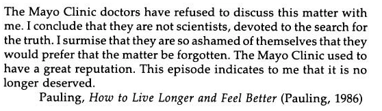
“The Mayo Clinic doctors refused to discuss these issues with me. I think they are not scientists, they are not dedicated to the search for truth. I think they are ashamed of themselves and prefer to forget about the issue. The Mayo clinic used to have considerable prestige. However, this process has shown me that it does not deserve it anymore.”
Pauling, How to Live Long and Feel Good (Pauling 1986)
However, Pauling’s efforts at the age of 80 paved the way for many studies on Vitamin C. Work continued in Japan, Germany and other countries.
In a study published at the end of 2015, Scientist Lewis Cantly showed that Vitamin C enters the cell and selectively kills the tumor, especially in intestinal cancer tumors with mutant KRAS and BRAF genes. Immediately afterwards, in the studies of Dr Allen and his friends from the University of Iowa in April 2017, all the mechanisms of action of Vitamin C were revealed very clearly. More importantly, the American Cancer Institute, which has been against Pauling for years, donated $9.7 million to the University of Iowa to support Vitamin C thousand cancer studies.
Vitamin C is very tightly controlled as it is absorbed from the intestines. Since it is very tightly controlled, the amount passed into your blood is very low even if 1gr is taken orally or 1 sack is eaten. At this concentration, the effect of Vitamin C shows antioxidant properties. In tumors, doctors do not want to use antioxidants. Because the tumor constantly produces acid, oxidation is at a high level. Clearing this oxidation can facilitate the work of the tumor.
Vitamin C is a pro-oxidant when administered intravenously. That is, hydrogen peroxide reverts to a state that produces oxygenated water. If we consider hydrogen peroxide as a powerful cleaning agent; We can compare it to bleach. More interestingly, the hydrogen peroxide produced by vitamin C is selectively produced only around tumor cells and has a direct toxic effect on the tumor. It only kills cancer cells; does not kill normal cells. For this reason, high-dose Vitamin C intravenously means that we are no longer just talking about a vitamin, but a pro-drug. Vitamin C administered intravenously in pharmacological doses is a prodrug.
The first reason why Vitamin C can distinguish a normal cell from a cancerous cell is the enzyme Catalase found in our blood and tissues. The Catalase Enzyme quickly converts hydrogen peroxide, that is, oxygenated water, into water and oxygen. However, this enzyme is almost absent in cancer cells. For this reason, cancer cells cannot remove hydrogen peroxide from the environment like normal cells.
The second reason is that when metals and hydrogen peroxide combine, they directly form free oxygen radicals, attacking and killing tumor DNA. In normal cells, the iron molecule is less than in cancer cells. Iron is one of the elements that ensures the survival of cancer stem cells. Increased iron combined with hydrogen peroxide selectively kills cancer cells.
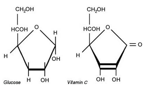
Vitamin C is a very interesting molecule. It is 90-95% similar to sugar. If blood sugar is measured in people taking vitamin C, the rate is high, because many machines cannot distinguish between sugar and vitamin C. Cancer cells do not use oxygen; It feeds on sugar alone, using 200 times more sugar than a normal cell. Therefore, when Vitamin C is given intravenously, it enters into cancer cells just like sugar.
You can think of it in the same way as the technical working system of PET CT/MR, which is taken to detect cancerous cells. Fluorine is attached to sugar to draw PET Film. The fluorine we bind is like a signal that indicates the location of the cancer. When you give fluorinated sugar intravenously, all the sugar is attracted by the cancer cells and shines in the film because the fluorine is bound. And those glowing cells are diagnosed as tumors. Therefore, Vitamin C has a very important role in cancer.
Even at very high doses, Vitamin C is excreted in the urine, as it is a water-soluble vitamin. There are only two rules:
If these rules apply, it can be said that Vitamin C has almost no side effects. The desired dose can be applied to the cancer patient by the doctor. There is no need for high doses for a healthy person. The purpose of administering high doses to cancer patients is that the pharmacological dose creates hydrogen peroxide in the blood.
However, some patients may experience discomfort when the Vitamin C concentration rises in intravenous intake. For this reason, a test dose of 15 g should be applied first. It should not be thought of as an allergy test to see side effects. To set the starting dose only, the reference point is set. Then the dose of Vitamin C is gradually increased. For example, if the target dose is 100gr, 100gr is not applied directly. First start with 25gr. Then it is increased to 50gr, 75gr and 100gr. Target doses above 50 g are definitely not started in the first application.
Vitamin C has a thirst-quenching effect as it increases urination. For this reason, it is recommended that patients drink water before the application. In addition, during the Vitamin C infusion, patients are definitely given oxygen. Thus, by increasing the free oxygen radicals formed inside, Vitamin C is provided to be more effective.
High-dose vitamin C products have been approved in Canada, Chile, and New Zealand since 2010. In addition, 25 gr/50 ml vials were approved by the FDA in the USA in 2017. In FDA reports, it is stated that it is safe up to a dose of 187 g/day.

Even more successful results are obtained when vitamin C is used with chemotherapy agents in the treatment of cancer patients. Vitamin C enhances the effect of chemotherapy. There are also some chemotherapy drugs that cannot be used. The reason for this is again explained by the functioning of the kidneys. Vitamin C also reduces the side effects of chemotherapy agents with which it can be used.
Especially in some types of cancer, smart drugs are used. For example, smart drugs are mostly preferred in bowel cancers. By looking at the EGFR pathway, by looking at the KRAS gene, effective drugs are selected accordingly. If these drugs are applied together with Vitamin C, much better results can be obtained.
Riordan Clinic is one of the clinics that performs Vitamin C application most frequently and has the highest number of cases. But the study of Dr Allen at the University of Iowa has revealed all the working principles of Vitamin C as well as showing all the mechanisms of action. Intravenous high-dose Vitamin C administration has proven to be safe and effective in both lung and brain tumors, along with chemotherapy and radiotherapy in cancer patients. When both phase 1 and phase 2 results of the studies were published, they were widely covered in medical journals.
In the state of Kansas, high-dose vitamin C therapy is administered at both the Riordan Clinic and the University of Kansas. The University of Kansas has a Vitamin C Infusion Clinic. One of the prominent names of this hospital, Dr. Levin, together with the head of the Department of Integrative Medicine at the University of Kansas, showed that Vitamin C enhances the treatment effects of chemotherapy. As a result of these studies, 25 g Vitamin C preparations were approved by the FDA in November 2017 and started to be used under license in the USA.
In the studies conducted by Samsung Hospital in South Korea on Vitamin C, it was revealed that when healthy people are treated with certain periods, they get sick less, they recover faster and their work efficiency increases. For this reason, especially in America, some countries of Europe, and South Korea, intravenous vitamin C is administered in non-serious viral infections and infections and at certain periods. It is a treatment that can be taken in cases of chronic fatigue. Besides its health effect, Vitamin C is ergogenic; gives energy. It also helps release endorphins. It makes the person feel good.
In chemistry there are Fenton reactions. Fenton reaction in many areas where chemistry serves; It is used in many fields such as textile and industry.
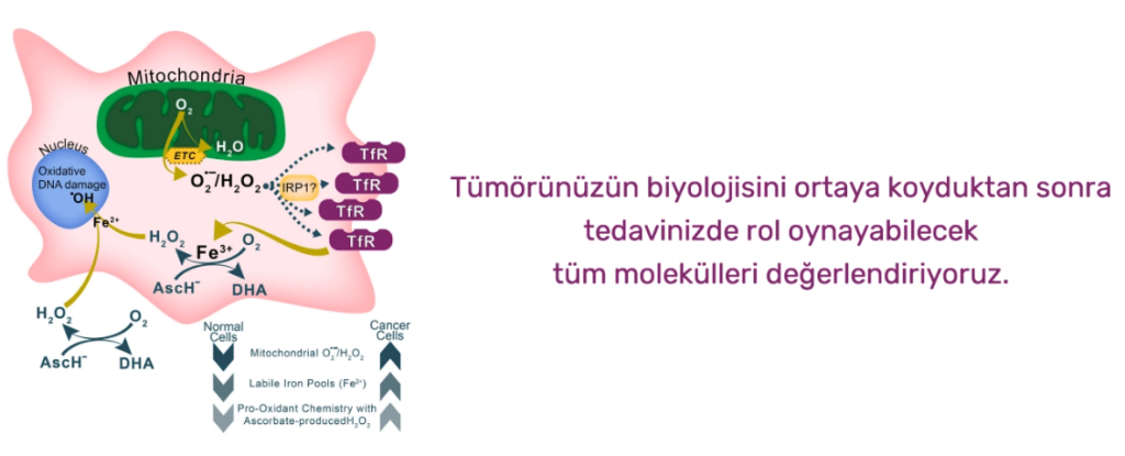
The basic mechanism is hydrogen peroxide, H2O, iron +2, iron +3 conversion in the environment, with the electron it gives off, and hydroxyl radicals form. They are also used in whitening and cleaning. When we look at the relationship with Vitamin C; ascorbic acid, namely Vitamin C, is the only molecule in nature that can donate 2 electrons. When it gives this electron, Iron +3 turns into Iron +2. And while this converted Iron +2 turns into Iron +3 again, the hydrogen peroxide formed by ascorbic acid in the environment provides the formation of hydroxyl radicals in the environment. In other words, Vitamin C first forms hydrogen peroxide and then hydroxyl radicals.
Hydrogen peroxide can also occur in our normal tissues. But it quickly turns it into water and oxygen with our existing enzymes. Since enzymes called catalase are not present in cancerous tissues, cancerous tissues cannot remove hydrogen peroxide formed by Vitamin C ascorbic acid. As a result, when you are infused, ascorbic acid donates its electron and turns into hydroxy ascorbate, while iron +3, iron +2 conversion hydroxy radicals are formed.
The excess iron molecules in cancer cells cause more interaction with hydrogen peroxide. Since the enzyme catalase is also deficient, the hydroxyl radicals formed selectively affect cancer cells. This is also called intravenous C Redox Chemotherapy due to reduction.
In summary, Fenton Reaction;
· During the conversion of ascorbic acid to dihydroxy ascorbate, hydrogen peroxide is formed in the presence of oxygen.
Because Vitamin C is very similar to sugar, it enters through the sugar channel. The same transformation occurs inside the cancer cell.
Together with the increased iron pool called ‘labile iron pool’, the hydroxyl radicals formed directly attack DNA and mitecontries and selectively kill cancer cells.
Vitamin C therapy selectively creates free oxygen radicals in cancer cells and causes damage to cancerous cells.
In the April issue of 2017’s Cancer Cell Journal, Dr. The study of Allen et al. has examined this issue in detail.
How Is Intravenous (Intravenous) Vitamin C Administered?
In particular, the infusion set must be protected from light when administering Vitamin C. A special colored set is used for this. In addition, since Vitamin C is a vitamin that deteriorates very quickly when left exposed, the IV bag must be protected from light.
The effect of Vitamin C lasts about 24 hours. It strengthens the immune system, especially against antiviral infection. It gives a better state of well-being the next day of application. In case of illness, it accelerates the healing process.
The main purpose of applying much higher doses when performing oncological treatments is to benefit directly from the tumor-killing effect, rather than to strengthen the immune system.
Excess vitamin C is not retained in the body, but is excreted through the urine. Pure Vitamin C does not smell in the urine.
Paraben-containing products can be used in applications that are said to be given as high-dose Vitamin C in wellness centers operating under the name of “Well Being”. When administering high doses of vitamin C, you may also be exposed to high doses of parabens.
The residence time of Vitamin C in the blood does not exceed 8 hours. Even if higher doses are given, this process only takes a little longer. If you really took Vitamin C, you would feel the need to urinate immediately.

Doctors make every effort to ensure that women with breast cancer can return to their usual activities as soon as possible. The recovery period varies for each woman, depending on the stage of the disease, the type of treatment and other factors.
After the operation, the woman’s arm and shoulder exercise helps this region regain strength and movement; reduces pain and muscle stiffness in the back and neck. Special exercises usually start a few days after the tubes that are placed under the skin and drained during the surgery and drain the accumulated blood and lymph fluid are removed. Exercises start slowly and for short periods, and can be done even in bed. It is mostly performed under the supervision of an experienced physiotherapist. As time passes, the quality and duration of the exercises can be increased. Regular exercise turns into a woman’s daily routine in the following period.
Women who have had a mastectomy or breast reconstruction should do the specific exercises their doctor will explain.
Doing exercises frequently and placing the arm on the pillow prevents or reduces lymphedema (swelling in the hand, arm and forearm) that may occur after surgery. Information about prevention and treatment of lymphedema is explained in the “Side Effects” section.
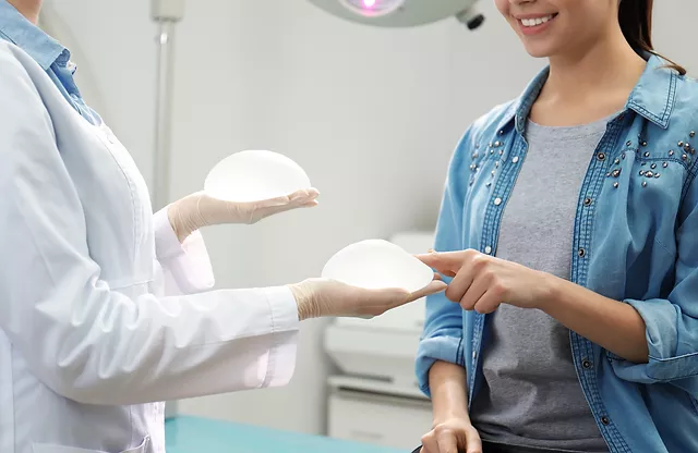
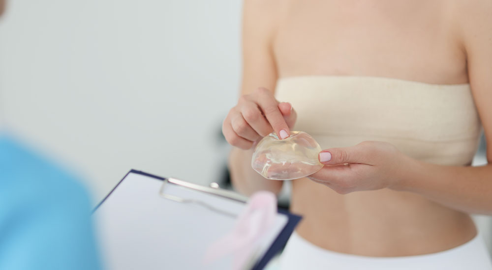
Silicone breast prostheses were created in 1963, inspired by the plastic bags used in blood transfusions that resemble a breast when carried. Silicone prostheses are materials filled with gel-like silicone, surrounded by a more solid silicone sheath, and can be produced in a round or oval shape. The consistency of the gel is very similar to that of an adult female breast and they are not noticeable on the body.
Silicon is one of the most common elements on earth. The combination of silicon with other elements creates a family of silicons with different structures. Depending on the properties of the joint, silicones can be found in solid, gel or liquid form (Figure 1). The most important difference between silicones used in industry and daily life and those used in breast prostheses is that medical silicones are pure and compatible with the body. Apart from breast prostheses, medical silicones are widely used in areas ranging from catheters to joint prostheses, heart valves and penile prostheses.
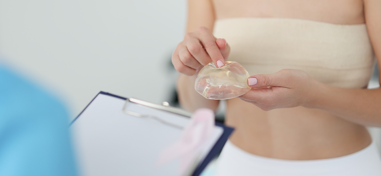
Figure 1. As the chains around the silicones lengthen and bond together, their consistency hardens
There are also silicone breast prostheses filled with water, not silicone gel. However, an implant filled with water is not preferred because it is harder in consistency. There is also the possibility of leakage and leakage in water-filled prostheses.
Types of Silicone Prostheses
Types of Implants by Shape:
Silicone prostheses are produced in drops and round forms. Drop prostheses are also called anatomical prostheses because their downward-expanding structure resembles the side view of the breast when viewed from the side (Figure 1).
Figure 1. Anatomical prostheses (top) and round prostheses (bottom)
Types of Implants According to the Consistency of the Gel It Contains:
Silicone prostheses are also divided into two according to the consistency of the gel they contain: soft gel and cohesive gel prostheses that keep their shape. Although soft gel implants are more suitable for the natural consistency of the breast, they are slightly displaced by gravity in their sheath. Cohesive gels, on the other hand, maintain their shape in any case and can not only enlarge the breast in volume, but also shape it into a certain shape. However, the breasts on which these prostheses are placed feel harder to the hand. In the last few years, slightly softer versions of Cohesive silicones have also been introduced as an option (Allergan® SoftTouch, etc.).
Most gel-containing dentures today use a form of gel that does not flow out when burst or cut (Fig. 2). This feature is a precaution to reduce the problems in leakages that may occur due to wear or accident.
Figure 2. An implant containing a cohesive gel does not flow out when burst or cut
If an implant containing a cohesive gel is used, the shape of the chosen silicone prosthesis will affect the shape of the enlarged breast. Which prosthesis to choose depends on what kind of breast shape you like. In general, women’s preferences about breasts can be divided into two (Figure 3):
● Rounded breasts that fill the décolleté and look like they’ve been pushed up with a bra
● Drop-shaped breasts that come with a flat slope down the décolleté and take a round form after the nipple
Figure 3. Which breast shape do you like more? It can be thought that those who prefer the two breast silhouettes on the left like drops, those who prefer the two silhouettes on the left like the round breast shape.
Although it is not a general rule, anatomical prostheses are preferred to achieve the drop shape, and round prostheses are preferred to achieve the round shape.
Other reasons for choosing anatomical prostheses are:
● In very thin patients with no breasts, a cohesive and rounded prosthesis can create a sudden and unnatural transition in the upper part. In such patients, using submuscular and anatomical prosthesis is a better option.
● In patients with slightly drooping nipples, the need for breast augmentation surgery can be reduced by using an anatomical prosthesis to raise the nipple.
The situations in which round prostheses are preferred are as follows:
● Patients who do intense sports such as tennis and swimming and who will be implanted under the muscle: If an anatomical prosthesis is used in these patients, the possibility of the prosthesis turning outward with muscle movements may create asymmetry. Even if round prostheses rotate, this may not produce a noticeable result.
● To fill the gap in the upper part of patients who will be augmented with breast augmentation.
The latest development in silicone technology is the implants containing gel in two different consistencies in the same sheath. Aiming for a more natural look, this technology is available in Polytech 4inTwo Series and Allergan’s 510 series. The part of the drop prosthesis that rests on the chest wall contains a softer silicone and the part under the nipple contains a harder silicone (Figure 4). Thus, the transition line created by the top part of the silicone, especially in the side profile, is eliminated and the projection of the nipple is increased.
Figure 4. Polytech® 4inTwo prostheses with two different densities of silicone
Implant Types According to Sheath Properties:
While the surface of the silicone sheath was flat and smooth in the first periods, textured forms were adopted later on (Figure 5). The sleeves of silicones are also technologically advanced and have become more sealed by adding several layers. The purpose of these changes is to reduce the event called capsular contracture. One of the rough forms is polyurethane coated prostheses. Once the silicone enters the body, this coating begins to melt and thus creates an intermediate zone that will reduce the formation of capsular contracture. It is especially preferred in patients who develop capsule contracture and whose prosthesis will be replaced.
Figure 5. Silicone prostheses with different sheath properties: Top left – formerly flat-surfaced transparent prostheses; Top right – polyurethane coated prosthesis; Bottom row – dentures with rough surface
Every woman’s breast is as unique as her fingerprint. The structure of the rib cage, its angle, the thickness and flexibility of the skin, the posture, the shape and width of the area where the mammary gland begins (footprint) are different in every woman and lead to the development of a personalized breast structure. The silicone prostheses produced, on the other hand, come in certain molds because they are fabricated. A manufacturer’s silicone variety is between 250-300. The most important step of breast augmentation surgery is to choose the most suitable prosthesis among them.
How to choose the most suitable prosthesis for you? In this regard, the first step is to decide what size you want. Psychological studies show that women do not dare to express the dimension they actually want. Another mistake made in this regard, especially in Turkish patients, is to confuse the size of the bra, such as 80-90, with breast size. The best expression of breast size is the “cup sizes” that go as A, B, C, D DD.
The next step is to choose the breast shape. You can do this by bringing photos of the breasts you like. At this stage, it becomes somewhat clear whether a round or drop-shaped prosthesis will be chosen.
Of course, you will also need to have a chest structure in which what you want can be applied. For this, your doctor takes detailed measurements of your breasts. These are listed as width, height, projection, amount of stretching, distance between neck and nipple, skin thickness, etc. The most critical of these measurements is the width of the breast. The limiting step in determining the size of the prosthesis is the width of the breast base. Because if you go sideways, more armpits will be entered, so it cannot be exceeded.
Then, your doctor chooses a few prostheses that are most suitable for you from the catalogs of companies that produce various silicone prostheses according to the measurements they take. There are two methods to pre-determine which of these will yield results: Your pre-operative photographs can be uploaded to the simulation system, and how the results will be obtained with the selected prostheses can be simulated. Crisalix® is one of these systems. By looking at this simulation, a sketch is formed that you can discuss with your doctor about what size or shape you want in general, although it is not exactly one-to-one. But the real live trial is done by placing the prostheses selected during the surgery on your chest and watching how you sit at the table while you sleep. Usually, several types of prostheses and trial pairs are brought in sizes close to the measurements made for the surgery.

It is the process of reconstructing breast tissue removed for cancer or other reasons. It is possible to make reconstructions that are very similar to other breasts with the developing techniques.
Studies and our own experiences show that breast reconstruction surgeries provide psychological support to women.
Thanks to the new technologies in medicine, the breast created in reconstruction surgeries can be very similar to the natural breast.
Almost all mastectomy patients can be suitable candidates for breast reconstruction. In these patients, breast reconstruction (repair) can be performed at the same time as mastectomy (breast removal). However, some patients may be advised by their surgeons to wait for reconstruction surgery (for example, if the breast is to be repaired with the patient’s own tissue (flap transfer), obesity, high blood pressure and smoking).
It can be observed after all surgical operations; The risks of bleeding, edema/fluid collection or anesthesia problems can also be observed after breast reconstruction surgeries.
In smokers, wound healing may be delayed or scarring may appear more. If a prosthesis is to be used, there may be a rare risk of developing an infection. In such cases, it may sometimes be necessary to remove the prosthesis and put it back in months later.
Capsular contracture is the most common problem in prosthesis use. In capsule cantracture; The scar tissue around the prosthesis compresses the prosthesis, which creates the feeling of breast hardness. Capsule contracture can be treated.
Breast reconstruction (repair) has no effect on cancer recurrence (recurrence). In addition, it does not create a situation that prevents radiotherapy or chemotherapy.
The outer part of all breast prostheses is made of silicone. Inside, there is silicone gel or saline (salt water mixture).
These two types of prostheses can also be used safely.

Onco-Aesthetic breast surgery; It means that surgery for breast cancer is performed together with plastic surgery methods to improve the aesthetic appearance of the breast. The method consists of combining the principles of oncological surgery and plastic surgery. The patient has both cancer surgery and the breast can be protected aesthetically.
Until the last decades, the entire breast was removed in breast cancer surgeries. Negative results were seen in terms of body integrity perception and psychological effects. Over the years, when it was understood that breast cancer could be treated without removing the breast, Onco-Aesthetic methods came to the fore.
Today, Onco-Aesthetic breast surgery primarily consists of removing large enough tissue to provide safe surgery without compromising the principles of cancer surgery in people who will undergo breast-conserving surgery, and closing the remaining breast space in the most aesthetic way. In addition, in patients with large breasts, it is possible to both get rid of cancer and reach a more suitable breast volume by reducing the breast with cancer and the opposite breast in the same session.
In patients for whom breast-conserving surgery is not possible, surgery can also be performed, if appropriate, by removing the inside of the breast completely and filling it with a silicone prosthesis or the person’s own tissues. Prostheses are used more frequently since they are now understood to be reliable.
Breasts can be reconstructed using the back muscles in those with a suitable body structure, and abdominal muscles and skin in those with a belly, and tummy tuck surgery is performed at the same time.
In patients with familial cancer risk or high-risk patients, prophylactic mastectomy can be performed without cancer by removing the inside of the breast and placing a prosthesis. Thus, the risk of breast cancer is minimized.
The method of Onco-Aesthetic surgery varies according to the patient, the type of treatment of the disease and time. The surgeon and the patient should discuss and choose the method together.
The advantages of Onco-Aesthetic surgery are that it allows wider surgery, better local control, better aesthetic results, reduced mastectomy rate, convenience to radiotherapy, providing breast equality, application in a single session, high quality of life, high patient satisfaction, cheaper and easier than late repairs.

Breast cancer can now be treated with developing surgical techniques without removing the entire breast. In this surgical treatment, cancerous tissue is removed with the onco-aesthetic surgical methods applied and the breast gains an upright appearance with breast aesthetics. With this method, there is a better view of the enlarged, deformed and even sagging breast tissue.
With the oncoesthetic surgery technique, the cancerous tissue is removed and then an aesthetic and upright breast can be made with the remaining breast tissue. Some patients also have this procedure done on their healthy breasts, depending on their wishes. Under the leadership of these developing new surgical techniques, patients diagnosed with cancer are somewhat motivated by the sense of renewal given by the plastic surgery to be performed. Oncoesthetic surgery techniques can be applied to both breasts. Optionally, it can be applied not only to the cancerous breast, but also to the healthy breast. After this procedure, it becomes upright, symmetrical and more aesthetic.
PET CT, which is considered one of the most effective imaging techniques today, is used in the diagnosis, staging, evaluation of response to treatment and radiotherapy planning processes of many cancers, especially lung, colon, head and neck cancers and lymphomas. PET CT has a great role in determining the epilepsy focus and investigating the presence of viable tissue in the heart after a heart attack. PET CT is an imaging technique that combines the powers of PET (Positron Emission Tomography) and CT (Computerized Tomography), which can provide detailed anatomical information, showing the functions of organs and tissues in the human body at the metabolic level. PET CT, which is used especially in the field of oncology, to detect the tumor, to determine the tumor grade, to evaluate the response to treatment, and in some cases to determine whether the existing mass is benign or malignant, has been widely used in our country as well as all over the world in the last 2 years.
PET CT examination method is most commonly used for oncological purposes such as investigating malignancy in known masses, staging of tumors, post-treatment follow-up, evaluation and early detection of recurrences, determination of response to treatment, staging of the primary tumor and determination of the course of the tumor, and determination of the biopsy site in a known mass.
Most cancers are highly curable if diagnosed early. For example, the 5-year survival rate in patients with early detection of breast cancer is over 80%. In many cancers, it is also very important to determine whether there is spread to the surrounding tissues or lymph nodes, that is, whether there is metastasis. Because one of the most important facts in the treatment of cancer is how it spreads to the organs in the body. Since it is possible to see the whole body at the same time in the images obtained with PET CT, it can also be evaluated whether the disease has spread to another organ.